|
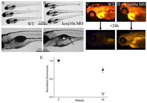
kcnj10a morphants have kidney defects. (A,B) The upper images shows three 120-hpf WT larvae (A) and three kcnj10a morphants (B), some of which have pericardial edema. The lower image is an enlargement of the swim bladder and pronephric duct area. In morphants, the pronephric duct is visible because it is dilated (dotted arrow) and, although partially obscured by pigment, the swim bladder is not visible (arrowhead). (C,D) The upper and lower images show a 72-hpf WT (C) and a kcnj10a morphant (D) immediately after fluorescent dextran injection (upper image) and 24 hours later (lower image). Although baseline fluorescence immediately after injection was comparable in WT and morphant, there was significantly higher remaining fluorescence after 24 hours in morphant (D) compared with WT (C) ZF. (E) Graph of measured fluorescence in WT (triangles; n=9) and morphant ZF (squares; n=12). y-axis shows fluorescence intensity normalised for the baseline measurement in WT. x-axis shows time (hours).
|