
Fig. 1
- ID
- ZDB-FIG-130719-2
- Publication
- Riera et al., 2013 - CERKL Knockdown Causes Retinal Degeneration in Zebrafish
- Other Figures
- All Figure Page
- Back to All Figure Page
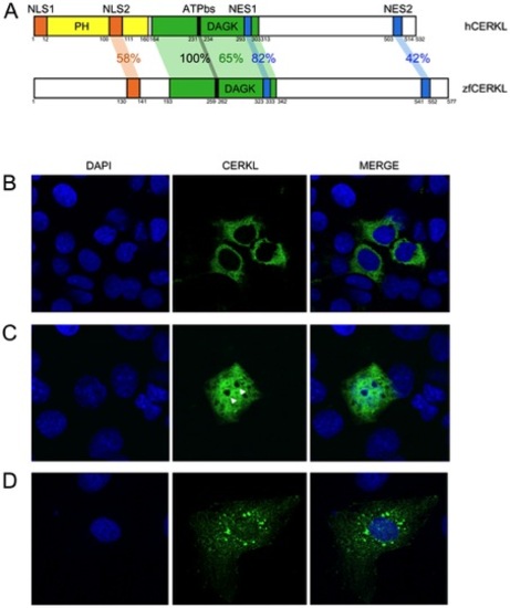
Human and zebrafish CERKL protein domains. (A) The reported hCERKL (NP_963842) protein domains described by either sequence homology (PH, pleckstrin homology; DAGK, diacylglycerol kinase and ATPbs, ATP binding site domains) or by functional analysis (NLS, nuclear localization signals; NES, nuclear export signals) and their conservation in zebrafish is shown in percentage of identity. (B–D) ZFCerkl-HA shares with human CERKL the dynamic subcellular localization in COS-7 transfected cells, shifting from the cytoplasm to the nucleus. Nuclei were stained with DAPI. Images correspond to individual optical sections. Photographs were at×63 magnification. (B) In most cells, ZFCerkl shows a uniform distribution in the cytosol and is absent from the nucleus. (C) Some cells per field showed localization of Cerkl in both, the cytosol and the nucleus, with clear exclusion from the nucleoli (white arrow). (D) Rarely, Cerkl contributes to cytosolic aggregates. |