
Fig. 6
- ID
- ZDB-FIG-130625-44
- Publication
- Schmid et al., 2013 - Loss of ALS-associated TDP-43 in zebrafish causes muscle degeneration, vascular dysfunction, and reduced motor neuron axon outgrowth
- Other Figures
- All Figure Page
- Back to All Figure Page
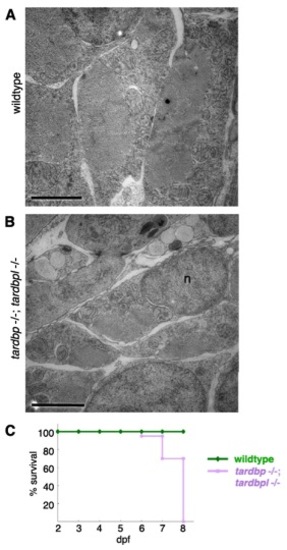
Severely degenerated myocytes of tardbp-/-;tardbpl-/- mutants. (A) Antibody staining of 2-dpf-old wild-type and tardbp-/-;tardbpl-/- mutant embryos with the myosin specific antibody ZE-BO-1F4 (green) and DAPI (blue). White arrowheads indicate degenerated myocytes. (B) Antibody staining of 1.5-dpf-old wild-type and tardbp-/-;tardbpl-/- mutant embryos with α-actinin (green), vinculin (red), and DAPI (blue). (A and B) Anterior to the left, lateral view. (Scale bars, 20 μm.) (C) EM pictures of skeletal muscle of a tardbp-/-;tardbpl-/- mutant embryo shows a highly disorganized pattern of thinner myofibrils (f) with disorganized network of sarcoplasmic reticulum (open arrowheads). Large part of the sarcoplasmic reticulum is misaligned and dilated (filled arrowheads). Individual myofibrils are not well separated from another and mitochondria can now be found within the muscle fibers at (black arrow). (2 dpf) (Scale bars, 500 nm.) |