
Fig. 4
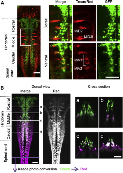
V2a Neurons that Project to the Spinal Cord(A) Texas red-Dextran was backfilled into a compound transgenic fish of Tg[chx10:Gal4] and Tg[UAS:GFP] at 3 dpf. Several ipsilaterally projecting large reticulospinal neurons, including MiD2i, MiD3i, MiV1, and MiV2 neurons, are GFP positive, showing that they are among the V2a neurons. The dots indicate neurons that are double positive for Texas red and GFP. (B) Kaede protein in the spinal cord of Kaede-expressing fish (Tg[chx10:Gal4] and Tg[UAS:Kaede] at 3 dpf) was photo converted by application of violet light to the spinal cord. After photo-converted Kaede (from green to red) was transported from the axons to the somata (8 hr waiting time), the observation was made. Green shows the distribution of green Kaede, whereas magenta shows the distribution of red (photo-converted) Kaede. The majority of V2a neurons in the caudal hindbrain are positive for Red-Kaede. In the more rostral region, the number of Red-Kaede-labeled neurons gradually became smaller. Subpanels of (a)–(d) show cross-sections. Scale bar represents 50 μm. |