FIGURE

Fig. 2

ID
ZDB-FIG-130524-3
Publication
Ramos-Balderas et al., 2013 - The zebrafish mutants for the V-ATPase subunits d, ac45, E, H and c and their variable pigment dilution phenotype
Other Figures
All Figure Page
Back to All Figure Page
Fig. 2

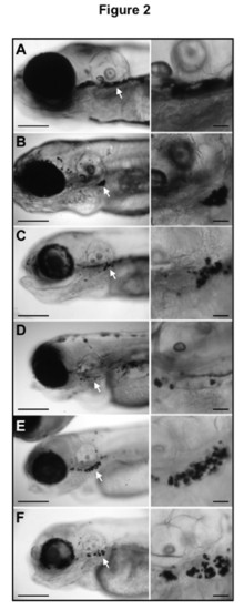
Lateral views from zebrafish insertional mutants for five V-ATPase subunits. Larvae WT (A) and mutants in subunits V0-d1 (B), V0-ac45b (C), V1-E1b (D), V1-H (E) and V0-ca (F) are shown. To the right from every image there is a close up view of the region indicated by the white arrow in the left panel. Melanin round spots accumulate ventrolateral to the ear, which is an identical feature observed in the class VI.C of zebrafish mutants from the Tubingen chemical screening (see reference 38). Even though the reason for this phenotypic characteristic is unknown, it could be used as a form to identify this type of mutations.

Expression Data

Expression Detail
Antibody Labeling
Phenotype Data
Fish:
Observed In:
Stage: Day 5

Phenotype Detail
Acknowledgments
This image is the copyrighted work of the attributed author or publisher, and ZFIN has permission only to display this image to its users. Additional permissions should be obtained from the applicable author or publisher of the image. Full text @ BMC Res. Notes