
Fig. S1
- ID
- ZDB-FIG-130524-13
- Publication
- Chou et al., 2013 - Fibronectin mediates correct positioning of the interrenal organ in zebrafish
- Other Figures
- All Figure Page
- Back to All Figure Page
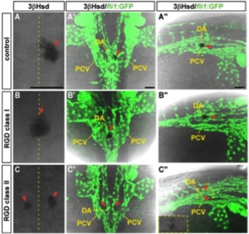
The effect of RGD peptide treatment on the interrenal tissue and the peri-interrenal vasculature. Sets of confocal images display the interrenal tissue as detected by 3β-Hsd activity staining (red arrows), and the midtrunk vasculature by fli1 promoter-driven green fluorescence, of 32-hpf Tg(fli1:EGFP)y1 embryos as treated with RGD peptide from 16 to 32 hpf. A–C, A′′–C′′: Dorsal views showing the midtrunk of the representative embryo for each phenotypic class, with anterior oriented to the top. A′′–C′′: Dorso-lateral views of the same embryo with anterior to the right. The interrenal region in C′′ is outlined, with its enlarged bright field image shown in the inset. The interrenal tissue in RGD-treated embryos displays affected laterality in the class I phenotype, and disrupted midline fusion in the class II phenotype. DA, dorsal aorta. PCV, posterior cardinal vein. Scale bar = 50 μM. |