
Fig. 6
- ID
- ZDB-FIG-130502-25
- Publication
- Song et al., 2013 - Pou5f1-dependent EGF expression controls e-cadherin endocytosis, cell adhesion, and zebrafish epiboly movements
- Other Figures
- All Figure Page
- Back to All Figure Page
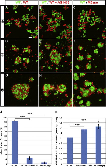
Pou5f1 and EGF Signaling Control Blastoderm Cell Cohesiveness(A–I) Primary cocultures of dissociated blastoderm cells from embryos labeled by microinjection of Alexa 488-dextran (green) or Rhodamine-dextran (red) at one-cell stage. (A–C) Dissociated cells shown 1 hr after plating, (D–F) after 4 hr incubation, and (G–I) after 8 hr incubation. Scale bar, 50 μm.(J) Percentage of clusters with intermingled cells out of total number of clusters (p < 0.001; WT/WT n = 23, WT/WT+AG1478 n = 36, and WT/MZspg n = 23 focal planes from four experiments).(K) Cell ratios in each cluster after 8 hr incubation (p < 0.001; number of clusters: WT/WT n = 49, WT/WT+AG1478 n = 131, and WT/MZspg n = 50).Error bars show SEM. |
Reprinted from Developmental Cell, 24(5), Song, S., Eckerle, S., Onichtchouk, D., Marrs, J.A., Nitschke, R., and Driever, W., Pou5f1-dependent EGF expression controls e-cadherin endocytosis, cell adhesion, and zebrafish epiboly movements, 486-501, Copyright (2013) with permission from Elsevier. Full text @ Dev. Cell