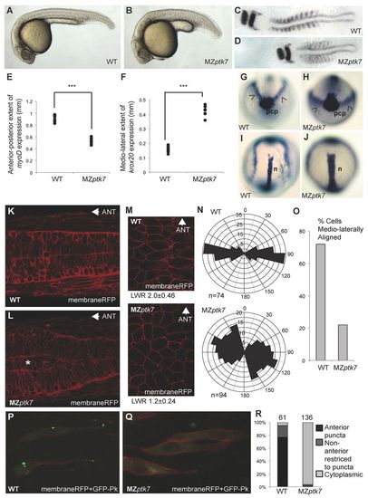
MZptk7 mutant embryos display PCP-mediated morphogenesis defects. (A,B) Lateral views of wild-type (A) and MZptk7hsc9 mutant (B) embryos at 24 hpf. (C,D) Flat-mounts of 10- to12 somite-stage wild-type (C) and MZptk7hsc9 (D) embryos stained for krox20 (hindbrain) and myoD (somite) gene expression. (E) Quantification of the anterior-posterior extent of the myoD expression domain in wild-type (WT) versus MZptk7hsc9 mutant embryos (***P<0.001, n=8 for each group). (F) Quantification of the mediolateral extent of krox20 expression in WT versus MZptk7hsc9 mutant embryos (***P<0.001, n=8 for each group). MZptk7hsc9 mutant embryos display clear defects in axial extension. (G-J) Anterior (G,H) and dorsal (I,J) views of bud stage WT (G,I) and MZptk7hsc9 (H,J) embryos stained for hgg1 (prechordal plate, pcp), dlx3 (prospective neural plate, arrowheads) and ntl (prospective notochord, n). MZptk7hsc9 mutants demonstrate defects in the convergence of both neuroectoderm and axial mesoderm tissues. (K,L) Dorsal confocal images of the neural tube and adjacent somites of 24 hpf wild-type (K) and MZptk7hsc9 (L) embryos, injected with membrane-localised monomeric RFP (membraneRFP) (Megason and Fraser, 2003). MZptk7hsc9 mutant embryos display an accumulation of neural progenitors (asterisk in L) at the centre of the neural primordium. Anterior is left. (M) MembraneRFP-labelled cells in the dorsal ectoderm of WT and MZptk7hsc9 embryos at 90% epiboly. Dorsal view, midline to the right and anterior to the top. The length-to-width (LWR) ratio of cells are as indicated for WT (n=74) and MZptk7hsc9 (n=94). (N) Rose diagrams for cell orientation relative to the embryonic midline at 90% epiboly in WT and MZptk7hsc9 embryos. (O) Graph showing percentage of mediolaterally aligned cells, for which longitudinal axis is oriented ±15° with respect to the embryonic mediolateral axis. (P,Q) Dorsal confocal images of the neural keel and adjacent somites of 8- to 10-somite-stage WT (P) and MZptk7hsc9 (Q) embryos scatter-labelled with GFP-Prickle (GFP-Pk) and membrane RFP. The subcellular localisation of the PCP marker GFP-Pk is disrupted in MZptk7hsc9. Anterior is up. Confocal imaging was carried out at the level of the first to the fifth somite pairs. (R) Quantification of the localisation of GFP-Pk puncta in WT (four embryos) versus MZptk7hsc9 (six embryos).
|
