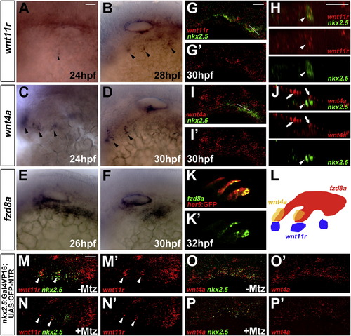
Expression of wnt11r, wnt4a, and fzd8a during Pouch Formation(A–F) Colorimetric in situs show expression of wnt11r in discrete domains of mesoderm (arrowheads in A and B), wnt4a in ectodermal patches (arrowheads in C and D), and fzd8a in pouch-forming endoderm (E and F).(G–J) Double fluorescent in situs show colocalization of wnt11r but not wnt4a with nkx2.5 in lateral views (G and I) and higher magnification orthogonal sections (H and J, taken at level of white lines in G and I). Arrowheads indicate mesoderm and arrows ectoderm.(K) Fluorescent in situ shows colocalization of fzd8a (green) with her5-positive pouch endoderm labeled by GFP immunohistochemistry (red).(L) Schematic showing expression of wnt11r in mesoderm (blue), wnt4a in ectoderm (yellow), and fzd8a in endoderm (red) during pouch formation.(M–P) Compared to un-injected siblings (M and O), injection of 5 nl of 5 mM Mtz into nkx2.5:Gal4VP16; UAS:CFP-NTR embryos results in reduced numbers of mesodermal cells expressing nkx2.5 and wnt11r (arrowheads), as well as reduced ectodermal wnt4a expression. Scale bars, 20 μM.
|
