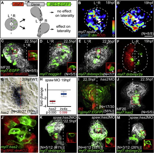
Tissue-Autonomous Inhibition of Bmp Activity Affects Cardiac Laterality(A) Cardiac laterality was assayed at 22.5 hpf in embryos with predominantly right-sided myocardial cell clones at 18 hpf.(B and B′) Spaw-expressing clones (false-colored white, indicated by asterisks) cause reversal of Bmp reporter Tg[BRE-AAVmlp:dmKO2]mw40 relative expression intensities, indicated by color range as shown for 18 hpf (B) and for 19 hpf (B′). Cardiac field is outlined by white dotted circle.(C) Control clones expressing enhanced green fluorescent protein (EGFP) do not affect cardiac jogging. Inset shows expression of the Tg[BRE-AAVmlp:dmKO2]mw40 Bmp reporter in control clone (asterisk) and neighboring cells.(D and E) Misexpression of the Bmp antagonist Noggin1 (D) or of dnBmpr2a (E) on the right side of the cardiac cone causes rightward cardiac jogging. Insets show specific suppression of Bmp activity within EGFP-positive misexpression clones (asterisk).(F) Fluorescence two-color in situ hybridization reveals normal lefty1 cardiac expression in clones expressing dnBmpr2a.(G) L/R asymmetric expression of has2.(H) Whisker box plots indicate relative expression changes of has2 and irx4a based on RT-qPCR analysis of cardiac cDNA (also see Supplemental Experimental Procedures for details on the statistical analysis). Unchanged expression of the cardiac stage marker irx4a shows that observed changes in has2 expression are not due to developmental delay.(I) Fluorescence two-color in situ hybridization of a has2 morphant with loss of cardiac laterality but correct lefty2 expression.(J) Clonal misexpression of Has2 on the right side of the cardiac cone affects cardiac laterality.(J2) Bmp activity is specifically suppressed within EGFP-positive Has2-misexpression clones (asterisks) but not in neighboring WT cells.(K–M) Cardiac laterality in spaw;has2 double morphants that have EGFP-positive dnBmpr2a misexpression clones. Cardiac laterality at 22.5 hpf strictly corresponds to the L/R distribution of dnBmpr2a-expressing cell clones. Shown are examples for mainly left-sided (K), evenly distributed (L), or right-sided (M) dnBmpr2a-expressing cell clones. Insets show that Bmp activity is specifically suppressed within EGFP-positive misexpression clones.L, left; R, right. Dotted line indicates the embryonic midline. Total number of embryos and percentiles indicate the occurrence of the most frequent expression patterns and phenotypes as shown. Scale bars, 50 μm.See also Movie S2.
|
