
FIGURE
Fig. 4
- ID
- ZDB-FIG-130409-25
- Publication
- Guner-Ataman et al., 2013 - Zebrafish second heart field development relies on progenitor specification in anterior lateral plate mesoderm and nkx2.5 function
- Other Figures
- All Figure Page
- Back to All Figure Page
Fig. 4
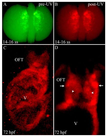
Photoconversion of nkx2.5+ cells in the ALPM reveals widespread contributions to SHF-derived structures. (A,B) Dorsal images of a 14- to 16-somite stage Tg(nkx2.5:Kaede) embryo before (A) and after (B) photoconversion with ultraviolet (UV) light. (C) Confocal z-stack image of the ventricle (V) and OFT at 72 hpf. (D) Confocal slice of the OFT. Arrowheads and arrows highlight endothelial and smooth muscle precursor cells in the OFT, respectively. Six out of eight photoconverted embryos exhibited red Kaede protein throughout the ventricle and OFT. ss, somite stage; OFT, outflow tract; hpf, hours post-fertilization. |
Expression Data
Expression Detail
Antibody Labeling
Phenotype Data
Phenotype Detail
Acknowledgments
This image is the copyrighted work of the attributed author or publisher, and
ZFIN has permission only to display this image to its users.
Additional permissions should be obtained from the applicable author or publisher of the image.
Full text @ Development