
Fig. 3
- ID
- ZDB-FIG-130404-15
- Publication
- Yoo et al., 2012 - The role of microtubules in neutrophil polarity and migration in live zebrafish
- Other Figures
- All Figure Page
- Back to All Figure Page
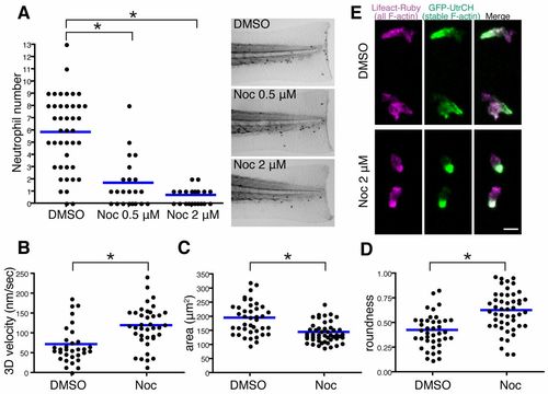
Microtubule depolymerization impairs neutrophil wound attraction but enhances neutrophil motility and F-actin polarity. (A) Neutrophil recruitment to wounded fins at 30minutes post wounding. Neutrophils were stained with Sudan Black. n = 42 (DMSO), 22 (Noc 0.5μM) and 22 (Noc 2&mu′M). (B–D) Microtubule depolymerization with nocodazole treatment enhances neutrophil 3D motility [(B), n = 32 (DMSO), n = 38 (Noc)], makes cells compact [(C), n = 40 (DMSO), n = 48 (Noc)] and induces a more round morphology [(D), n = 40 (DMSO), n = 48 (Noc)]. Tg(mpx:GFP-UtrCH) was used for live analysis. (E) Nocodazole treatment enhances polarity of F-actin dynamics in neutrophils. Rear localization of stable F-actin detected by GFP-UtrCH is particularly emphasized after microtubule depolymerization. Tg(mpx:GFP-UtrCH) was crossed with Tg(mpx:Lifeact-Ruby). *P<0.05, one-way ANOVA with Dunnett post-test (A) and two-tailed unpaired t-test (B–D). Data in A are representative of at least three separate experiments and three time-lapse movies were analyzed for data in B–D. Scale bar: 10 μm. |