|
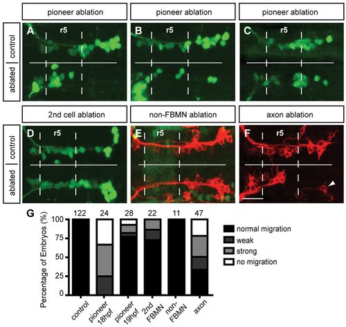
Pioneer neuron ablation blocks migration of follower FBMNs. (A-C) Maximum projection images of Tg(islet1:GFP) (green) embryos at 24 hpf, after ablation of the pioneer neuron on one side of the embryo at 18-19 hpf. (A) Pioneer neuron ablation blocks follower FBMN migration. (B) Strong partial migration phenotype results in two to four FBMNs reaching r6. (C) Weak partial migration phenotype results in migration of five to eight FBMNs into r6. (D) Ablation of the second FBMN to migrate does not affect FBMN migration in most embryos. (E) Tg(zCREST1:membRFP/pGFP5.3) embryos label FBMNs (red) and neuroepithelial cells in r5 (green), respectively. Ablation of a single r5 neuroepithelial cell in the path of FBMN migration does not affect migration. (F) Ablation of the trailing axon of the pioneer neuron, using Tg(zCREST1:membRFP) embryos, blocks FBMN migration; arrowhead indicates presumed pioneer neuron. (G) Percentage of embryos affected by ablation. Scale bar: 20 μm.
|