|
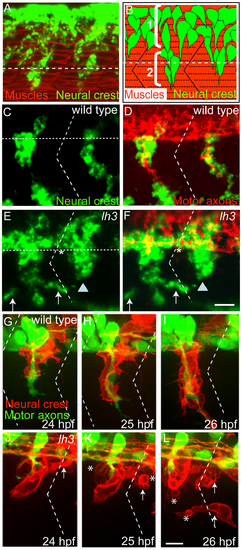
The role of lh3 signaling in neural crest cell migration. (A) Lateral view of a 22 hour old embryo stained with the pan-neural crest cell marker crestin (riboprobe, in green) marking neural crest cells, and with F59 antibody marking adaxial muscle cells. (B) Schematic diagram showing two modes of zebrafish neural crest cell migration: 1) sheet-like, non segmental and 2) stream-like, segmental. Neural crest cells (green) migrate through a central region of each somite after forming segmental streams (red). White horizontal dashed lines in A, B, C and E indicate approximate position of the ventral boundary of the neural tube. (C and D) Lateral view of a 28 hpf embryo stained with crestin (green) and the motor neuron maker antibodies (Znp1 and SV2; red). Horizontal dotted lines in A indicate the ventral boundary of the neural tube. E and F show lateral views of lh3 mutant embryos stained with crestin and motor neuron markers. Arrows in E and F point to neural crest cells near the somite boundaries. Asterisks marks neural crest cells forming sheet between two adjacent hemisegments. G–L shows still images from time lapse movies recorded from embryos expressing membrane bound RFP (Tg[sox10:mrfp]) in neural crest cells and GFP in motor neurons (Tg[mnx1:gfp]). In wildtype embryos (G–I), neural crest cells always migrate mid-segmentally along with motor axons. In lh3 mutant embryos (J–L), neural crest cells migrate along with motor axons mid-segmentally and in the region near segment/somite boundary (arrows). Neural crest cells in lh3 mutants also acquire a rounded morphology (asterisks). Oblique dotted lines in C to L indicate position of the somite/segment boundary. Scale bars:10 micron.
|