|
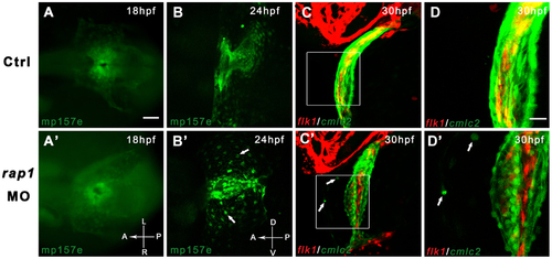
Time-lapsed observation revealed abnormal heart development in Rap1 knock-down zebrafish. Transgenic fish mp157e without any injection (A) and injected rap1MO (A′) were indistinguishable in early heart development. By 24 hpf, rap1MO injected mp157e embryos, crossed in flk1:mCherry background, started to show abnormality in heart development and remodeling (B to B′), including the failure of heart tube extension, abnormal cardiac looping and incomplete heart chamber formation. In higher magnification views, the morphants show that a small portion of GFP labeled cardiac cells (arrows in B′ to D′) did not migrate properly to be eventually packed in the heart tube. Dorsal view, head to left in A to C and A′ to C′. Lateral view, head to left in B to D and B′ to D′. Scale bar, 100 µm in A to C, and A′ to C′; 40 µm in D and D′.
|