
FIGURE
Fig. 1
- ID
- ZDB-FIG-130225-15
- Publication
- Feng et al., 2012 - The Stress-Response Gene redd1 Regulates Dorsoventral Patterning by Antagonizing Wnt/β-catenin Activity in Zebrafish
- Other Figures
- All Figure Page
- Back to All Figure Page
Fig. 1
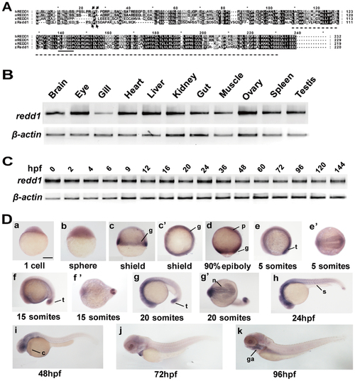
Zebrafish redd1 encodes a conserved protein and is expressed in many tissues. A) Alignment of REDD1/Redd1 sequence from human, mouse, Xenopus, and zebrafish. Conserved residues are shaded. The RTP801_C domain is marked by a dotted line. Arrows mark the two Thr residues critical for human REDD1 phosphorylation and degradation. The conserved 14-3-3 binding site is indicated by a solid line. B) RT-PCR analysis of the indicated adult tissues. C) RT-PCR analysis of zebrafish embryos at the indicated stages. hpf, hours post fertilization. D) Whole mount in situ hybridization analysis of zebrafish embryos at the indicated stages. (a–c, d) Lateral views with the animal pole oriented at the top; (c2) Top view from the animal pole. (e, f, g, h–k) Lateral views with the anterior oriented toward the left. (e2, f2, g2) Ventral views with the anterior oriented toward the left. c, common cardinal vein; g, germ ring; ga, gill arches; n, neural ectoderm; p, prechordal plate/mesoderm; s, somite; t, tail bud. Scale bar = 200 µm. |
Expression Data
| Gene: | |
|---|---|
| Fish: | |
| Anatomical Terms: | |
| Stage Range: | 1-cell to Adult |
Expression Detail
Antibody Labeling
Phenotype Data
Phenotype Detail
Acknowledgments
This image is the copyrighted work of the attributed author or publisher, and
ZFIN has permission only to display this image to its users.
Additional permissions should be obtained from the applicable author or publisher of the image.
Full text @ PLoS One