
Fig. 7
- ID
- ZDB-FIG-130212-17
- Publication
- Coffey et al., 2013 - Novel oxytocin gene expression in the hindbrain is induced by alcohol exposure: transgenic zebrafish enable visualization of sensitive neurons
- Other Figures
- All Figure Page
- Back to All Figure Page
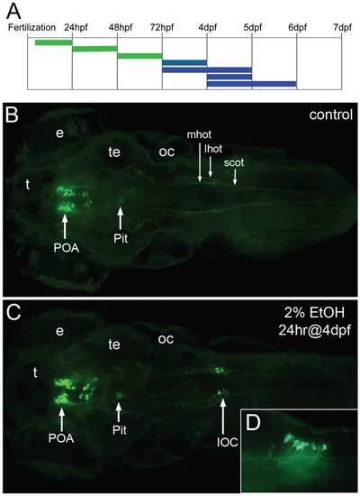
Expression of the oxtl:GFP transgene is induced in the hindbrain by exposure to ethanol. A, the duration and embryonic stage of 2% ethanol treatments are illustrated with solid bars. Oxtl:GFP expression in the neuroendocrine preoptic area was unaffected with all treatments (green), while oxtl:GFP was induced in the hindbrain by later treatments (blue). B, C, Tg(oxtl:GFP) larva shown at 6 dpf, dorsal views. B, control embryo, C, treated with 2% ethanol for 24 hrs at 4 dpf. A small group of large GFP-expressing neurons appears in the hindbrain of the ethanol-treated larva. We call these ethanol-induced GFP-expressing neurons, induced oxytocin cells (IOC). D, inset, a higher magnification view of the IOCs shows processes that appear to enter the hindbrain oxytocinergic tracks, lateral view. Eyes (e), lateral hindbrain oxytocinergic tract (lhot), medial hindbrain oxytocinergic tract (mhot), otic capsule (oc), pituitary (Pit), preoptic area (POA), spinal cord oxytocinergic tract (scot), tectum (te), telencephalon (t). |
| Gene: | |
|---|---|
| Fish: | |
| Condition: | |
| Anatomical Terms: | |
| Stage: | Day 6 |