
Fig. 3
- ID
- ZDB-FIG-130129-12
- Publication
- Folgueira et al., 2012 - Morphogenesis underlying the development of the everted teleost telencephalon
- Other Figures
- All Figure Page
- Back to All Figure Page
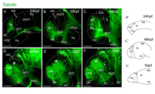
Neuropil organization in the telencephalon between 1 dpf and 5 dpf. A-F. Acetylated α-tubulin inmunostaining. The pallial telencephalic neuropil (indicated by asterisks) expands greatly posterior to the OB between days 2 and 5. The forebrain flexure places the subpallium and hypothalamus more posteriorly from 24 hpf to 36 hpf (A and B). At 2 dpf, the olfactory bulb neuropil is located close to the dorsal rim of the AIS and there is no obvious pallial neuropil. The pallial neuropil (asterisk) expands greatly posterior to the OB between days 2 and 5 (D-F). Lateral views, anterior to the left. A′, C′ and F′ illustrate the orientation of telencephalon in the related photomicrographs. Scale bars, 50 μm. AC: anterior commissure; AIS: anterior intraencephalic sulcus; Di: diencephalon; DVDT: dorsoventral diencephalic tract; E: epithalamus; Ha: habenula; Hy: hypothalamus; Me: mesencephalon; OB: olfactory bulb; OC: optic chiasm; ON: olfactory nerve; PC: posterior commissure; POC: postoptic commissure; SOT: supraoptic tract; TAC: tract of the anterior commissure; Tel: telencephalon; TPC: tract of the posterior commissure; TPOC: tract of the postoptic commissure. |