
Fig. S2
- ID
- ZDB-FIG-130123-12
- Publication
- Opitz et al., 2012 - Transgenic zebrafish illuminate the dynamics of thyroid morphogenesis and its relationship to cardiovascular development
- Other Figures
- All Figure Page
- Back to All Figure Page
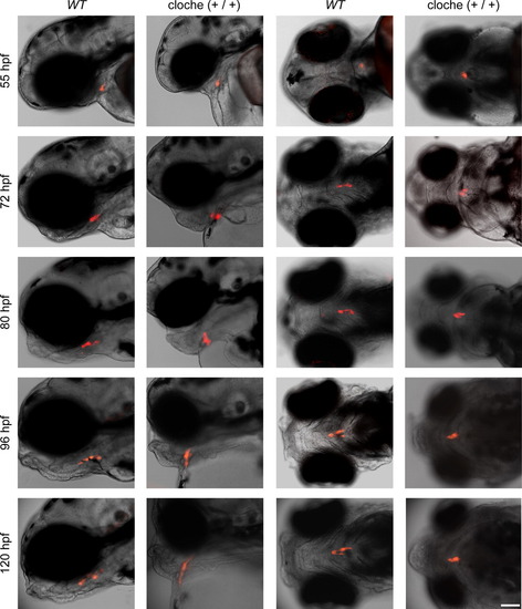
Live imaging of thyroid development in wild-type (WT) and cloche embryos expressing mCherry in thyroid cells. The two columns on the left show lateral views and the two columns on the right show ventral views of embryos that were transiently embedded in low melting agarose for live imaging at the indicated time points. The normal expansion of thyroid tissue along the pharyngeal midline seen in WT embryos was defective in cloche embryos. Note that the progressive increase of the pericardial edema caused a marked disruption of the normal morphogenesis of the hypobranchial region as a whole. Merged images from brightfield and epifluorescence microscopy are shown. Scale bar: 100 μM. |
Reprinted from Developmental Biology, 372(2), Opitz, R., Maquet, E., Huisken, J., Antonica, F., Trubiroha, A., Pottier, G., Janssens, V., and Costagliola, S., Transgenic zebrafish illuminate the dynamics of thyroid morphogenesis and its relationship to cardiovascular development, 203-216, Copyright (2012) with permission from Elsevier. Full text @ Dev. Biol.