
Fig. 5
- ID
- ZDB-FIG-130115-15
- Publication
- Mellman et al., 2012 - Fibrillin-2b regulates endocardial morphogenesis in zebrafish
- Other Figures
- All Figure Page
- Back to All Figure Page
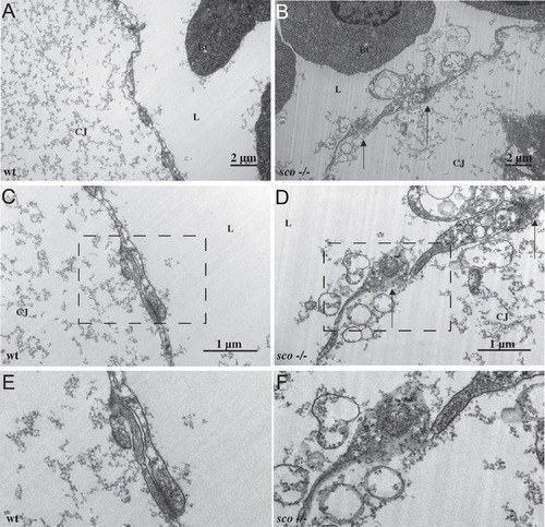
Electron microscopy study of endocardium details adhesion defects. Sections of 30 hpf wild-type (A), (C), (E) and scote382 mutants (B), (D), (F) at 9.4 kX (A) and (B) and 19 kX (C) and (D). Panels E and F correspond to the boxed areas of C and D, respectively. Wild-type endocardial cells (A), (C), (E) form a continuous layer between the cardiac jelly and the cardiac lumen. Mutant cells (B), (D), (F) fail to adhere to each other leading to a disorganized barrier, and allowing cardiac jelly to leak into the lumen. In (B), (D) and (F), the lumen and blood cells are located in the upper-left, while space between the endocardium and myocardium (which the cardiac jelly should occupy) is in the lower-right. Black arrows point to breaks between endocardial cells. CJ: cardiac jelly; BC: blood cell; L: lumen. |
| Fish: | |
|---|---|
| Observed In: | |
| Stage: | Prim-15 |
Reprinted from Developmental Biology, 372(1), Mellman, K., Huisken, J., Dinsmore, C., Hoppe, C., and Stainier, D.Y., Fibrillin-2b regulates endocardial morphogenesis in zebrafish, 111-119, Copyright (2012) with permission from Elsevier. Full text @ Dev. Biol.