
Fig. 2
- ID
- ZDB-FIG-130102-10
- Publication
- Delaune et al., 2012 - Single-cell-resolution imaging of the impact of notch signaling and mitosis on segmentation clock dynamics
- Other Figures
- All Figure Page
- Back to All Figure Page
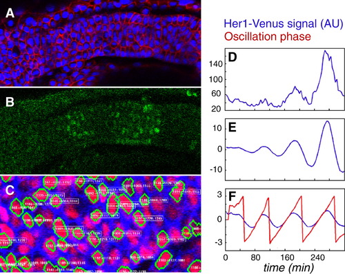
Detection and Analysis of Clock Oscillations at Single-Cell Resolution(A and B) Confocal section of the PSM of a 12-somite-stage heterozygous her1:her1-venus embryo (lateral view, anterior left) injected with membrane-mCherry and H2B-Cerulean mRNAs at the one-cell stage. Raw signals for membrane-Cherry (red) and H2B-Cerulean (blue) are shown in (A), and for Her1-Venus (green) in (B).(C) Image resulting from automated 3D segmentation of confocal pictures and cell tracking using our MATLAB-based program. Green cell contours are automatically generated and can be manually deleted or added. White and yellow numbers indicate tracking information, which is also automatically generated and can be manually corrected and validated.(D) Raw fluorescence data from a single PSM cell showing four oscillations over time.(E and F) Illustration of the heuristic algorithm used to compute the oscillation phase. First the signal is smoothed, then the average value over a predefined time window comparable to the period is removed (E), and finally the amplitude of the signal is rescaled over the same time window to obtain a pseudo sine wave (F, blue line). Phase (F, red line) is computed as detailed in the Experimental Procedures.See also Figure S3. |
Reprinted from Developmental Cell, 23(5), Delaune, E.A., François, P., Shih, N.P., and Amacher, S.L., Single-cell-resolution imaging of the impact of notch signaling and mitosis on segmentation clock dynamics, 995-1005, Copyright (2012) with permission from Elsevier. Full text @ Dev. Cell