
Fig. S2
- ID
- ZDB-FIG-121212-18
- Publication
- Dumortier et al., 2012 - Collective mesendoderm migration relies on an intrinsic directionality signal transmitted through cell contacts
- Other Figures
- All Figure Page
- Back to All Figure Page
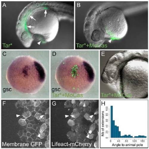
Activation of the Nodal pathway in the absence of casanova drives cells to a prechordal plate fate. (A and B) Injection of one cell at the 16-cell stage with RNAs encoding the constitutive form of the Nodal receptor (Tar*) leads cells to take part in endodermal derivatives (arrows) and hatching gland (arrowhead, A). When casanova expression is simultaneously blocked by a morpholino, injected cells take part in the hatching gland (B; n = 26 of 29 embryos). (C–H) Cells injected with Tar* RNAs and casanova morpholino were transplanted to the lateral margin of a host embryo at the onset of gastrulation. During gastrulation, these cells express the prechordal plate marker goosecoid (C and D), and take part in the hatching gland at 24 hours post-fertilization (E; n = 15 of 16 embryos). (F–H) During gastrulation, these cells migrate in group toward the animal pole (F–H; n = 19 of 21 embryos). Labeling their membrane and actin cytoskeleton (Lifeact) shows that they emit actin-rich extensions (white arrowheads), with the same frequency (0.216 extensions·cell1·min1) and orientation (H) as endogenous plate cells (P = 0.43, n = 357 and n = 537 extensions; compare Fig. S2H vs. Fig. 1L). Animal pole is to the top (C and D) or left (F and G). |