
Fig. S3
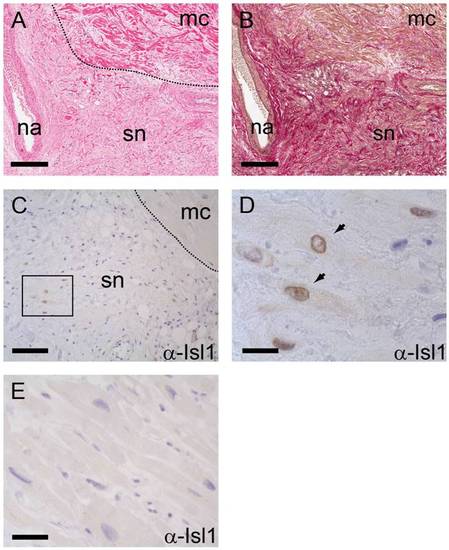
Immunohistochemical detection of Islet-1 in human cardiomyocytes in the sinoatrial node. (A) Hematoxylin and eosin staining of the sinoatrial node. SN indicates the area of the node showing where the specialized cardiomyocytes are located. MC indicates myocardium adjacent to the node. NA indicates the nodal artery. The boundary between SN and MC is highlighted by the dotted line. Scale bar represents 400 μm. (B) Elastic van Giesen stain of a consecutive section of (A) illustrating that the cardiomyocytes are embedded within collagen and elastic tissue. Scale bar represents 400 μm. (C) Islet-1 immunostain of sinoatrial node. SN indicates sinoatrial node. MC indicates myocardium adjacent to the node. The boundary between SN and MC is highlighted by the dotted line. Scale bar represents 160 μm. (D) Magnification of the boxed region in (C). Islet-1 immunostain with positive brown staining of the nuclei of the cardiomyocytes. On average 5% of the cardiomyocytes in the sinoatrial node revealed a positive signal. Scale bar represents 40 μm. (E) Staining is absent in the myocardium next to the sinoatrial node. Scale bar represents 80 μm. |