
Fig. 5
- ID
- ZDB-FIG-121031-27
- Publication
- Nadjar-Boger et al., 2012 - Structural and functional analysis of myostatin-2 promoter alleles from the marine fish Sparus aurata: Evidence for strong muscle-specific promoter activity and post-transcriptional regulation
- Other Figures
- All Figure Page
- Back to All Figure Page
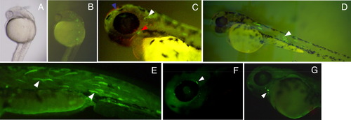
Transient expression analysis of saMSTN-2 promoter activity in zebrafish embryos. The DNA construct saMSTN-2b promoter (1324/22)/GFP was injected into zebrafish zygotes at 1-2 cell stages. GFP expression was analyzed at 24 and 48–50 hpf. A mosaic pattern of expression was observed. A, B. GFP in neural crest of 24 hpf embryos, shown in dorso-lateral view by direct visualization of live embryos under bright field (A) and fluorescence (B) stereoscope. C, 50 hpf live embryos showing GFP expression in otic epithelium (white arrowhead), forebrain (blue arrowhead) and near the developing head musculature (red arrowhead). D, GFP expression in myofibers at 48 hpf (live embryos). E–G. GFP immunodetected in 50 hpf embryos in myofibers (arrowheads, E), near the eye (arrowheads, F) and near the heart (arrowheads, G). (For interpretation of the references to color in this figure legend, the reader is referred to the web version of this article.) |
Reprinted from Molecular and Cellular Endocrinology, 361(1-2), Nadjar-Boger, E., Hinits, Y., and Funkenstein, B., Structural and functional analysis of myostatin-2 promoter alleles from the marine fish Sparus aurata: Evidence for strong muscle-specific promoter activity and post-transcriptional regulation, 51-68, Copyright (2012) with permission from Elsevier. Full text @ Mol. Cell. Endocrinol.