
Fig. 6
- ID
- ZDB-FIG-121023-39
- Publication
- Quick et al., 2012 - Expression analysis of zebrafish membrane type-2 matrix metalloproteinases during embryonic development
- Other Figures
- All Figure Page
- Back to All Figure Page
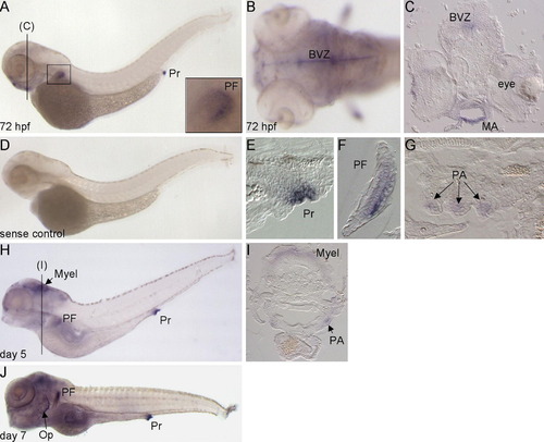
Expression pattern of mmp15b in larval stage embryos. (A and D) Lateral and dorsal (B) views of 72 hpf embryos with anterior to the left. Inset in panel (A) shows close-up of pectoral fin, PF. (C) Cross-section through the brain ventricular zone (BVZ) and mandibular arch (MA) of a 72 hpf embryo. (E–G) Sagittal plane sections of 72 hpf embryos showing mmp15b expression in the proctodeum (Pr), PF, and anterior pharyngeal arches, PA. (H and J) Lateral views of day 5 and 7 embryos with anterior to the left. (I) Cross-section through the myelencephalon (Myel) and PA of a day 5 embryo (position shown in panel H). Op, operculum. |
| Gene: | |
|---|---|
| Fish: | |
| Anatomical Terms: | |
| Stage Range: | Protruding-mouth to Days 7-13 |
Reprinted from Gene expression patterns : GEP, 12(7-8), Quick, R.E., Dunlap, J.A., and Jessen, J.R., Expression analysis of zebrafish membrane type-2 matrix metalloproteinases during embryonic development, 254-260, Copyright (2012) with permission from Elsevier. Full text @ Gene Expr. Patterns