
Fig. 5
- ID
- ZDB-FIG-120907-11
- Publication
- Xing et al., 2012 - Zebrafish foxP2 Zinc Finger Nuclease Mutant Has Normal Axon Pathfinding
- Other Figures
- All Figure Page
- Back to All Figure Page
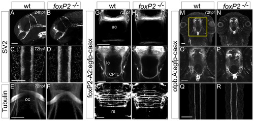
foxP2 does not affect axon pathfinding. Confocal z-stack images of whole-mount embryos, scale bars 50 μm, show no difference between wild-type and foxP2 mutant (/) embryos for axon pathfinding using a variety of axonal labels. (A–D) anti-SV2 immunohistochemistry at 24hpf, lateral views of the brain, rostral to the left (A, B) and 72hpf, dorsal views of the spinal cord, rostral to the top (C, D). (E, F) anti-acetylated tubulin immunohistochemistry at 72hpf, ventral views of the optic chiasm. (G-L) GFP immunohistochemistry at 72hpf in Tg(foxP2-enhancerA.2:egfp-caax) embryos that labels foxP2 neurons show no pathfinding errors in anterior commissure (ac), longitudinal commissures (lc), tract of the commissure of the posterior tuberculum (TCPTc), or reticulospinal axons (rs). (M-R) GFP immunohistochemistry at 72hpf in Tg(otpb.A:egfp-caax) embryos for visualization of dopaminergic and neuroendocrine projections (M, N, with insets shown in O, P) in the brain, and dopaminergic axon tracts in spinal cord (Q, R). |
| Gene: | |
|---|---|
| Antibody: | |
| Fish: | |
| Anatomical Terms: | |
| Stage Range: | Prim-5 to Protruding-mouth |