
FIGURE
Fig. S4
- ID
- ZDB-FIG-120810-42
- Publication
- Feng et al., 2012 - Live Imaging of Tumor Initiation in Zebrafish Larvae Reveals a Trophic Role for Leukocyte-Derived PGE(2)
- Other Figures
- All Figure Page
- Back to All Figure Page
Fig. S4
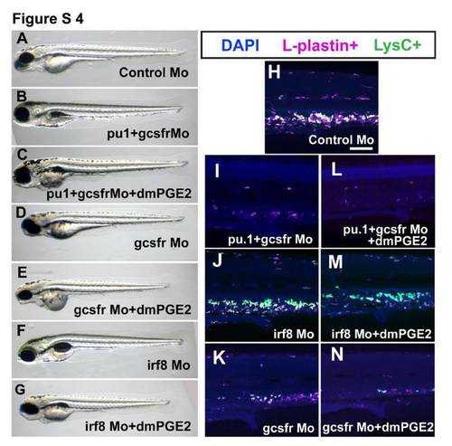
Normal morphology of leukocyte-depleted morphants (related to Figure 3) (A-G) Wide field images of morphants showing no general morphological defects; (H-N) Confocal images of anti-L-plastin immunostaining (magenta) of Lys:DsRed+ (green) larvae, showing no increase of L-plastin+ cells (magenta) or LysC:DsRed+ cells (green) in dmPGE2 treated, pu1+gcsfr morphants (L), irf8 morphant (M) or gcsfr morphants (N), when compared with untreated equivalent morphants (I, J, K respectively). Scale bar=100μm. |
Expression Data
| Genes: | |
|---|---|
| Fish: | |
| Anatomical Terms: | |
| Stage: | Day 4 |
Expression Detail
Antibody Labeling
Phenotype Data
| Fish: | |
|---|---|
| Condition: | |
| Knockdown Reagents: | |
| Observed In: | |
| Stage: | Day 4 |
Phenotype Detail
Acknowledgments
This image is the copyrighted work of the attributed author or publisher, and
ZFIN has permission only to display this image to its users.
Additional permissions should be obtained from the applicable author or publisher of the image.
Full text @ Curr. Biol.