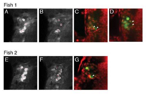
Laser ablation of single, identified, tangential nucleus neurons, (related to Figure 5).
Two examples showing targeted laser-ablation of tangential nucleus neurons. (a, e) Images of single z-planes after photoactivation of the tangential nucleus, with red targeting spots indicating locations near the center of selected cells where the pulsed infrared laser was spiral-scanned. (b, f) Images immediately after ablation showing disrupted cellular integrity caused by cavitation bubbles within the targeted neurons (red spots). Note that in some cases adjacent cells were photobleached but these do not show evidence of cavitation bubbles. (c, d, g) Confocal images after fixation and TO-PRO-3 staining (red). We were able to locate the autofluorescent signature of cavitation bubbles (which appear as green crescent-shaped structures, arrowheads). These lesion sites were associated with an absence of nuclear staining, whereas immediately adjacent cells displayed intact nuclei. (c) and (d) are two different z-planes, in the same specimen, in which we located the four cavitation bubbles corresponding to four ablated tangential neurons. Intact green cells, which do not have a crescent-shaped appearance, represent photoactivated neurons that were not targeted for ablation; these invariably have red nuclei. All images show dorsal views of the left tangential nucleus region, anterior top. Two examples are shown from a total of six larvae.
|
