
Fig. S4
- ID
- ZDB-FIG-120725-45
- Publication
- Vasilyev et al., 2012 - Mechanical Stretch and PI3K Signaling Link Cell Migration and Proliferation to Coordinate Epithelial Tubule Morphogenesis in the Zebrafish Pronephros
- Other Figures
- All Figure Page
- Back to All Figure Page
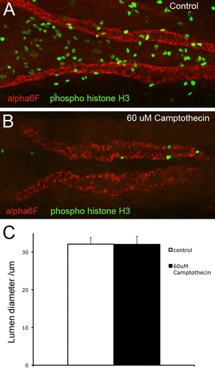
Obstruction-induced cell stretch does not depend on cell proliferation, related to Figure 2. 48 hpf ET33d10 fish were subjected to distal obstruction for 8hours, fixed and stained with anti- pospho histone H3 (green) and anti alpha6F (red) antibodies (A,B). 60 μM Camptothecin was used to inhibit cell proliferation (monitored by the amount of phospho histone H3 staining). Maximal luminal diameter of the proximal kidney was used as a measure of radial cell stretch and was identical in the experimental and the control condition (C). (A,B) show flattened confocal stacks. The apparent kidney staining with phospho histone H3 in (B) is actually in a different focal plane (in the CNS). No significant kidney phospho histone H3 staining is observed after 8hours of obstruction. |