
Fig. 5
- ID
- ZDB-FIG-120720-40
- Publication
- Zou et al., 2012 - Crb Apical Polarity Proteins Maintain Zebrafish Retinal Cone Mosaics via Intercellular Binding of Their Extracellular Domains
- Other Figures
- All Figure Page
- Back to All Figure Page
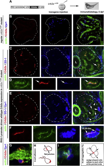
Transgenic Expression of Wild-Type Crb2a in crb2am289 Mutants Led to Photoreceptor Aggregation (A) A schematic illustrates the structure of the transgene construct that was injected into embryos at 1-4-cell stages to express wild-type Crb2a with the RH2-1 green opsin promoter in photoreceptors (Tsujimura et al., 2007). It should be noted that the number of transgenic photoreceptors varies from embryo to embryo because of varying efficiencies of transgenesis in the injected fish. (B) In regular crb2am289 mutant retinas, Zpr1-positive cones (blue) scatter everywhere in the retina; note the lack of Crb2a signals (red). (C and D) Retinal cells that transgenically expressed wild-type Crb2a (red) tended to form aggregates. The OLM-like structures, visualized by the OLM marker ZO-1 (arrows), were present in the aggregates. Actin staining (green) illustrated the overall structure of the retina as well as the OLM-like structures (D, arrows). (D) shows a cell aggregate in (C) (arrow) at a higher magnification. (E and F) The identical tissue section showed in (C) and (D) was subsequently immunostained with zpr1 antibodies; this confirmed that the aggregating cells were mostly double cones (F, higher magnification). (G and H) Two Crb2a-expressing transgenic cells established a flat junctional interface, which was highly enriched with Crb2a (arrows). (H) shows the contours of transgenic cells in (G). (I and J) Four wedge-shaped transgenic double cones adhered to each other to form a mini rosette, and Crb2a localized to the central region of the rosette (arrows). (J) shows the contours of transgenic cells in (I). Dashed lines in (B, C, and E) mark the boundary of the retinas, and the letters L indicate the lens. See also Figure S4. |
| Antibody: | |
|---|---|
| Fish: | |
| Stage: | Day 5 |
| Fish: | |
|---|---|
| Observed In: | |
| Stage: | Day 5 |
Reprinted from Developmental Cell, 22(6), Zou, J., Wang, X., and Wei, X., Crb Apical Polarity Proteins Maintain Zebrafish Retinal Cone Mosaics via Intercellular Binding of Their Extracellular Domains, 1261-1274, Copyright (2012) with permission from Elsevier. Full text @ Dev. Cell