
Fig. 5
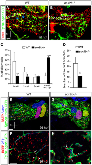
Intrahepatic biliary network morphogenesis and bile canaliculi formation are impaired in sox9b mutants. (A–B) Distribution of hepatocytes and biliary cells in 96 hpf wild-type and sox9b mutant larvae as revealed by Prox1 (red) and Tg(Tp1bglob:GFP) (green) expression. In wild-type, the cell bodies of biliary cells are separated from one another and interconnected via cellular processes (A). In sox9b mutants, the biliary cells are clustered together (B). Dashed lines mark the long bile ducts and arrows in (A) point to the short interconnecting ducts that are missing in the mutants. (C) Percentage (average±SEM) of biliary cells that exist as single cells, doubles, triples, or clusters of four or more at 96 hpf. 6 wild-type and 6 sox9b mutant larvae were analyzed. (D) Number (average±SEM) of branching points observed along the bile ducts per liver. 7 wild-type and 7 mutants were analyzed. (C–D) Asterisks indicate statistical significance: *p<0.05; ****p<0.0001; *****p<0.00001. (E, G) Wild-type and sox9b mutant livers stained for BSEP (red) which marks the canaliculi, Alcam (blue) which marks the apical side of the hepatocytes, and Tg(fabp10:ras-GFP) (green) which labels the hepatocytes. The hepatocytes in wild-type livers are organized in parallel arrays (highlighted by pseudo-colors), while the sox9b mutant hepatocytes are arranged in rosettes (highlighted by pseudo-colors). (F, H) High magnification confocal images showing the morphology of biliary cells and bile canaliculi. The canaliculi (arrows) in sox9b mutant livers appear shorter and wider compared to wild-type. (A–B) All images are projections of confocal z-stacks. (E–H) Single confocal plane images. (A–B, E–H) Ventral views, anterior (A) to the top. Scale bars, 20 μm. |
| Fish: | |
|---|---|
| Observed In: | |
| Stage: | Day 4 |