
FIGURE
Fig. 1
- ID
- ZDB-FIG-120627-18
- Publication
- Ton et al., 2012 - Semaphorin3d mediates Cx43-dependent phenotypes during fin regeneration
- Other Figures
- All Figure Page
- Back to All Figure Page
Fig. 1
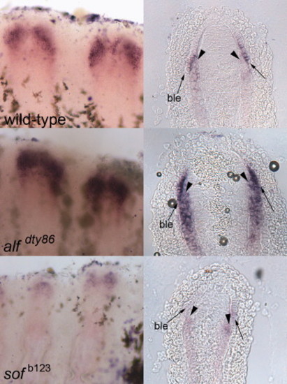
sema3d is differentially expressed in wild-type (top), alfdty86 (middle) and sofb123 (bottom). Left: whole mount in situ hybridization shows increased expression in alfdty86 and decreased expression in sofb123 compared to wild-type. Right: Cryosections reveal the tissue-specific localization of sema3d-expressing cells. Arrowheads point to skeletal precursor cells; arrows point to the basal layer of the epidermis (ble). |
Expression Data
| Gene: | |
|---|---|
| Fish: | |
| Condition: | |
| Anatomical Terms: | |
| Stage: | Adult |
Expression Detail
Antibody Labeling
Phenotype Data
Phenotype Detail
Acknowledgments
This image is the copyrighted work of the attributed author or publisher, and
ZFIN has permission only to display this image to its users.
Additional permissions should be obtained from the applicable author or publisher of the image.
Reprinted from Developmental Biology, 366(2), Ton, Q.V., and Iovine, M.K., Semaphorin3d mediates Cx43-dependent phenotypes during fin regeneration, 195-203, Copyright (2012) with permission from Elsevier. Full text @ Dev. Biol.