
Fig. 2
- ID
- ZDB-FIG-120612-2
- Publication
- Moro et al., 2012 - In vivo Wnt signaling tracing through a transgenic biosensor fish reveals novel activity domains
- Other Figures
- All Figure Page
- Back to All Figure Page
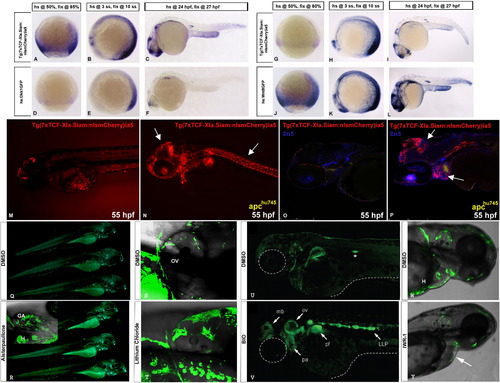
TCFSiam lines represent bona fide Wnt/β-catenin reporters. (A–F) In situ hybridization with an antisense mCherry probe on Tg(7xTCF-Xla.Siam:nlsmCherry)ia5/ Tg(hsp70l:dkk1-GFP)w32 embryos. In embryos heat-shocked at 50% epiboly, 3 somite-stage and 24 hpf, mCherry RNA is detected at 85% epiboly, 10 somite-stage and 27 hpf, respectively. (A–C) Heat-shocked Tg(7xTCF-Xla.Siam:nlsmCherry)ia5 single transgenic control embryos stained for mCherry RNA. (A) During late gastrula, reporter mRNA is expressed in a broad marginal domain. (B) During somitogenesis, Wnt/β-catenin activity is detectable in the midbrain-hindbrain boundary, and the posterior neuroectoderm and mesoderm. (C) During organogenesis, the reporter is expressed in multiple domains of the CNS, the lateral line primordium, the epidermis of the yolk extension and weakly in posterior tail. (D–F) In heterozygous Tg(7xTCF-Xla.Siam:nlsmCherry)ia5/Tg(hsp70l:dkk1-GFP)w32, mCherry signal is strongly reduced in all expression domains. (G–L) In situ hybridization with an antisense mCherry probe on Tg(hsp70l:wnt8a-GFP)w34/Tg(7xTCF-Xla.Siam:nlsmCherry)ia5 embryos. (G–I) Heat-shocked Tg(7xTCF-Xla.Siam:nlsmCherry)ia5 single transgenic control embryos stained for mCherry RNA. Note that the signal intensities in A-C and G-I are different due to variable duration of staining. (J–L) In heterozygous Tg(hsp70l:wnt8a-GFP) w34/Tg(7xTCF-Xla.Siam:nlsmCherry)ia5, a much stronger signal is detected. (M–P) Reporter activity in 55 hpf Tg(7xTCF-Xla.Siam:nlsmCherry)ia5 in apchu745 background (N and P). Wild type siblings (sib) are shown in M and O. Immunostaining for zn8 antibody is visible in blue. Tg(7xTCF-Xla.Siam:nlsmCherry)ia5 expression is upregulated ectopically in the heart, brain region and lateral region (arrows) according to a constitutive activation of the Wnt signaling pathway in apc mutants. (Q,R) Treatment for 36 h with 5 µM Alsterpaullone (R) increases Wnt reporter activity in the pharyngeal arches of 3 dpf Tg(7xTCF-Xla.Siam:GFP)ia4 larvae. Parallel treatment with control DMSO (Q) does not alter reporter activity. (S,T) Administration of 0.1 M LiCl for 36 h increases GFP fluorescence in the otic vesicle and pharyngeal arches (white arrowheads) of 3 dpf Tg(7xTCF-Xla.Siam:GFP)ia4 (T) when compared to age-matched transgenic larvae treated with DMSO (S). (U,V). Incubation of transgenic embryos in BIO from 24 hpf to 48 hpf, induces a robust tissue specific activation of the reporter (arrows in V). Notice that BIO treated animals resemble apc phenotype in lateral line, with an hypertrophic, stalled and strongly GFP expressing primordium, while in 2% DMSO control embryos the secondary lateral line primordium is already migrating (asterisk in U). (W,Y) Treatment of 24 hpf Tg(7xTCF-Xla.Siam:GFP)ia4 embryos with 10 µM IWR-1 for 24 h (Y) reduced reporter fluorescence particularly at the heart level (white arrow) when compared with DMSO treated embryos (W). ga: gill arches; h:heart, ov:otic vesicle. |
Reprinted from Developmental Biology, 366(2), Moro, E., Özhan, G., Mongera, A., Beis, D., Wierzbicki, C., Young, R.M., Bournele, D., Domenichini, A., Valdivia, L.E., Lum, L., Chen, C., Amatruda, J.F., Tiso, N., Weidinger, G., and Argenton, F., In vivo Wnt signaling tracing through a transgenic biosensor fish reveals novel activity domains, 327-340, Copyright (2012) with permission from Elsevier. Full text @ Dev. Biol.