
Fig. 3
- ID
- ZDB-FIG-120601-35
- Publication
- van Ham et al., 2012 - Apoptotic Cells Are Cleared by Directional Migration and elmo1- Dependent Macrophage Engulfment
- Other Figures
- All Figure Page
- Back to All Figure Page
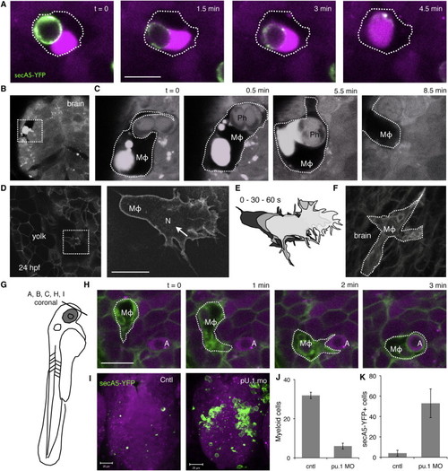
Primitive Macrophages Are Responsible for Engulfment(A) Engulfed secA5-YFP+ cell fuses with fluorescent vacuole in macrophage.(B and C) Large vacuole marked by red fluorescent protein originally expressed in neurons and dark surrounding shape indicates presence of macrophage in the forebrain (rostral view).(D) Image of memGFP transgenic animals showing a large migratory cell on yolk resembling a macrophage. Nuclear Fli1 GFP expression (arrow), a marker for primitive macrophages, is visible in the nucleus (N) of the presumptive macrophage.(E) Cartoon depicts dynamics of the cell.(F) memGFP labeled macrophage near the otic vesicle in the hindbrain at 24 hpf.(G) Drawing of 48 hpf larva, indicating orientation of coronal optical sections shown in (D)–(F), (H), and (I) [38].(H) Images showing a representative example of a macrophage in the forebrain engulfing an apoptotic cell. Nuclear condensation, characteristic of apoptosis, can be distinguished by the darker spot in fluorescently marked apoptotic cell (marked by “A”). Macrophages are indicated by “MΦ.”(I) WT (left, Cntl) and PU.1 morphant (right) brains are marked by transgenic expressed red fluorescent protein in secA5-YFP transgenic animals <60 hpf.(J) Quantification of macrophages on yolk by memGFP in control and PU.1 morpholino-injected animals.(K) Quantification of apoptotic cells in forebrain region shown in (I) in control and PU.1 morphants. Data are represented as mean ± SEM in (J) and (K); p < 0.05. Scale bars represent 10 μm (A, D, and H). Also see Figure S2. |