
Fig. 4
- ID
- ZDB-FIG-120525-28
- Publication
- Gao et al., 2012 - Dcc Regulates Asymmetric Outgrowth of Forebrain Neurons in Zebrafish
- Other Figures
- All Figure Page
- Back to All Figure Page
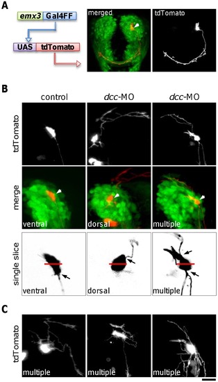
Inhibition of Dcc function causes ADt axons to project dorsally or to form multiple processes. (A) Labeling of individual ADt neurons by mosaic expression of fluorescent protein tdTomato. Image of a live 36 hpf Tg(lhx5BAC:Kaede) animal that was injected with emx3:Gal4FF and UAS:tdTomato plasmids is shown. The tdTomato labeled neuron projected an axon into the AC. Merge panel shows the position of the tdTomato labeled soma (marked by an arrowhead). Scale bar = 50 μm. (B) Injection of dcc morpholino causes ADt neurons to project axons dorsally or to form multiple processes. Labeled ADt neurons are marked by arrowheads in the merged panels. Left panels show an ADt neuron with a normal ventrally projecting axon in a control animal. Middle panels show an ADt neuron with an aberrant dorsally projecting axon in a dcc-MO injected animal. Right panels show an ADt neuron with both ventrally and dorsally projecting processes. Black arrow in the single slice images indicates the origin of the axon on the surface of the cell body. Red bar indicates the middle of the dorsal and the ventral side of the labeled neuron cell body. Scale bar equals to 20 μm in the projected images or 10 μm in the single slice images. (C) Additional examples of ADt neurons with multiple aberrant axons in dcc-MO injected animals. Scale bar = 15 μm. |
| Fish: | |
|---|---|
| Knockdown Reagent: | |
| Observed In: | |
| Stage: | Prim-25 |