
Fig. S1
- ID
- ZDB-FIG-120508-41
- Publication
- Fiaz et al., 2012 - Swim-Training Changes the Spatio-Temporal Dynamics of Skeletogenesis in Zebrafish Larvae (Danio rerio)
- Other Figures
- All Figure Page
- Back to All Figure Page
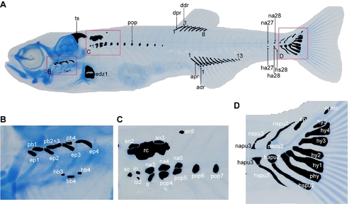
The cartilage structures, indicated here in black, which were analyzed in this study. A) Alcian blue staining of 14 dpf trained fish, lateral view. The areas indicated in red are shown enlarged in B, C and D. adr, anal distal radial; apr, anal proximal radial; ddr, dorsal distal radials; dpr, dorsal proximal radial; edz1, endoskeletal disc with cartilage subdivision zone 1; ha27/28, haemal arch 27/28; hs, haemal spine 28; na27/28, neural arch 27/28; pop, paraphophysis; ts, posterior end of the tectum synoticum. B) Branchial structures, lateral view. bb, basibranchial; ep, epibranchial; hb, hypobranchial; pb, pharyngobranchial. C) Weberian apparatus, lateral view. in, intercalarium; lp2, lateral process 2; na, neural arch; pop, paraphophysis; rc, roofing cartilage; sc, scaphium; sn, supraneural; tr, tripus. D) Caudal fin, lateral view. ep, epural; hapu, haemal arch of preural; hspu, haemal spine of preural; hy, hypural; napu, neural arch of preural; nspu, neural spine of preural; opstc, opistural cartilage; phy, parhypural. Nomenclature follows Cubbage and Mabee [5], Bird and Mabee [6] and Bensimon-Brito et al. [45]. |