
Fig. 3
- ID
- ZDB-FIG-120508-38
- Publication
- Yin et al., 2012 - Regulation of zebrafish heart regeneration by miR-133
- Other Figures
- All Figure Page
- Back to All Figure Page
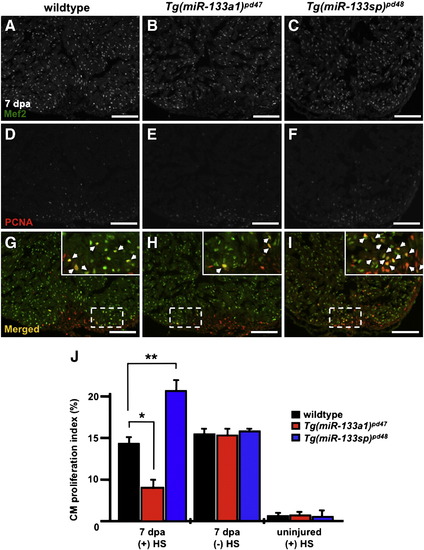
miR-133 restricts cardiomyocyte proliferation during heart regeneration. A–I) Wildtype, Tg(miR-133a1)pd47 and Tg(miR-133sp)pd48 adult hearts were resected, allowed to regenerate for 6 days, and subjected to a single heat-treatment at 38 °C. Representative 7 dpa wildtype, Tg(miR-133a1)pd47 and Tg(miR-133sp)pd48 heart sections were stained with Mef2 (A–C) and PCNA (D–F) and subsequently merged (G–I). Insets in (G–I), high zoom images of the white dashed rectangle; arrowheads indicate proliferating CMs. J) CM proliferation indices were determined for each group at 7 dpa. n = 10–12; Mean ± SEM, Student′s t-test p-value < 0.001 for * and **. HS = heat-shock. Scale bar in (A–I) represents 100 μm. |
Reprinted from Developmental Biology, 365(2), Yin, V.P., Lepilina, A., Smith, A., and Poss, K.D., Regulation of zebrafish heart regeneration by miR-133, 319-327, Copyright (2012) with permission from Elsevier. Full text @ Dev. Biol.