
Fig. 4
- ID
- ZDB-FIG-120427-15
- Publication
- Singh et al., 2012 - Regeneration of amputated zebrafish fin rays from de novo osteoblasts
- Other Figures
- All Figure Page
- Back to All Figure Page
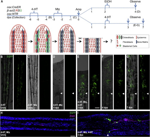
New Osteoblasts Arise in the Regenerates of Osteoblast-Depleted Fins through De Novo Differentiation (A) Cartoon depicting strategy that combines inducible lineage tracing and cell ablation in the osx:CreER; β-act2:RSG; osx:NTR background. 4-HT treatment imparts a β-actin2-driven EGFP label in osteoblasts. (B and C) 4-HT labels osteoblasts of uninjured fins with EGFP fluorescence (B). This label was undetectable after Mtz treatment (C), indicating efficient osteoblast ablation. (D) After amputation of fins of 4-HT-labeled and Mtz-treated animals, EGFP label was not detectable in 4 dpa regenerates or portions of the fins proximal to the injury site. Because the label was driven by the β-actin2 promoter, this result indicates that EGFP loss in (C) was due to cell ablation and not downregulation of an osteoblast marker. (E–G) A second 4-HT treatment at 2 dpa generated EGFP+ cells in 3, 4, and 7 dpa regenerates, but not in portions of the fins proximal to the injury site. This result indicates that, although β-actin2 expressing osteoblasts are not contributed by uninjured fin regions, the regenerated osteoblasts can still be labeled by EGFP via their expression of osx:CreER and β-act2:RSG after amputation. (H) Longitudinal section of uninjured fin corresponding to (C), indicating lack of EGFP fluorescence after 4-HT labeling and subsequent Mtz treatment. (I) Longitudinal section of 3 dpa regenerate corresponding to (E), indicating that EGFP fluorescence induced by a postamputation 4-HT label is present in the osteoblast compartment of the regenerated portion only. Scale bar = 100 μm. See Figure S4. |
Reprinted from Developmental Cell, 22(4), Singh, S.P., Holdway, J.E., and Poss, K.D., Regeneration of amputated zebrafish fin rays from de novo osteoblasts, 879-886, Copyright (2012) with permission from Elsevier. Full text @ Dev. Cell