
Fig. 8
- ID
- ZDB-FIG-120405-49
- Publication
- Cavaco Rodrigues et al., 2012 - Skeletal muscle regeneration in Xenopus tadpoles and zebrafish larvae
- Other Figures
- All Figure Page
- Back to All Figure Page
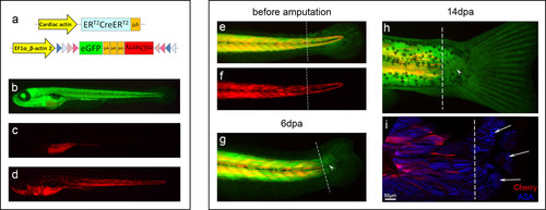
Labelled myofibres do not contribute to new muscle during regeneration of zebrafish larvae tail. (a) Representation of the Car-ERCreER and eab2-EGFPTmCherry [52] constructs, which allow the expression of mCherry in muscle fibres after activation of Cre. (b) These transgenics expressed eGFP ubiquitously. (c) Without tamoxifen treatment, mCherry expression was not observed (gut is autofluorescent). (d) After tamoxifen treatment, mCherry was generally visible in many muscle fibres. (e, f) Tails of two weeks old zebrafish larvae were amputated proximal to the base of the fin fold. (g) Six days later, some animals had regenerated a small piece of tail (arrowhead) and a caudal fin. (h) At 14 dpa, the small tail regenerate (arrowhead) continued to be free of mCherry labelling. (i) Confocal image of the same region, showing that the small tail regenerate has muscle fibres (arrows, ASA: α-sarcomeric actin) that are not labelled with mCherry. Pictures e to i correspond to the same animal. Dashed line: amputation plane. n = 33. |