
Fig. S4
- ID
- ZDB-FIG-120330-23
- Publication
- Zhao et al., 2012 - Kinesin-2 family in vertebrate ciliogenesis
- Other Figures
- All Figure Page
- Back to All Figure Page
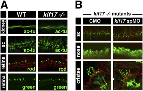
kif17 mutant phenotype. (A) Top two rows show confocal images of whole embryos stained with anti-acetylated tubulin antibody (green) to visualize cilia in the pronephric duct and the spinal canal (sc) at 30 hpf. Bottom two rows show confocal images of transverse cryosections through the retina at 5 dpf stained with anti-rod opsin or anti-green opsin antibodies (green). (B) Confocal images of kif17 mutant larvae at 3 dpf treated with control morpholino (CMO) or anti-splice kif17 morpholino as indicated. Larvae were stained with anti-acetylated tubulin antibody (green) to visualize cilia in the spinal canal, nasal pit, and ear cristae. When shown, red channel contains phalloidin staining. |
| Fish: | |
|---|---|
| Knockdown Reagent: | |
| Observed In: | |
| Stage Range: | Prim-15 to Day 5 |