
Fig. 4
- ID
- ZDB-FIG-120329-44
- Publication
- Gomez et al., 2012 - Identification of Vascular and Hematopoietic Genes Downstream of etsrp by Deep Sequencing in Zebrafish
- Other Figures
- All Figure Page
- Back to All Figure Page
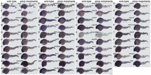
Analysis of gene expression in etsrp morphants. Flk1-gfp embryos were injected at the one cell stage with a mixture of translation blocking morpholinos for etsrp and analyzed by WISH at the 24hpf stage. Marked reduction is apparent in all genes examined in the axial vasculature, which includes the dorsal aorta and posterior cardinal vein, and is marked with a downward facing arrow in all images. The primitive myeloid cells stained in (L) myo1f wild-type controls are absent in their etsrp morphant counterparts. The staining in the axial trunk region of rgl2 in etsrp morphants marks primitive erythrocytes that are trapped due to lack of circulation. The expression of non-vascular structures is not affected otherwise in etsrp morphants. Embryos are positioned laterally with the anterior facing left. Abbreviations: cht, caudal hematopoietic tail region; cv, cranial vasculature; da, dorsal aorta. Scale bar: 250 μm. |