
Fig. 1
- ID
- ZDB-FIG-120315-61
- Publication
- Friedrich et al., 2012 - Mutation of zebrafish dihydrolipoamide branched-chain transacylase E2 results in motor dysfunction and models maple syrup urine disease
- Other Figures
- All Figure Page
- Back to All Figure Page
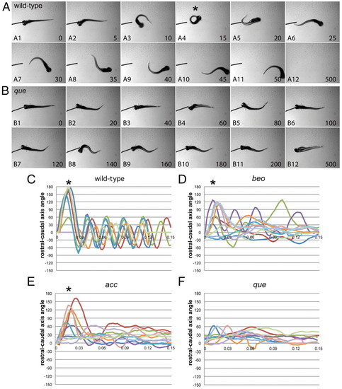
que mutants exhibit abnormal swimming behavior at 96 hpf. (A,B) Selected frames from high-speed video recordings are shown with times indicated in milliseconds. (A) A wild-type larva demonstrates a normal C-bend (A4, asterisk) in response to a touch stimulus, followed by smaller-amplitude body undulations to clear the field (A5-A12). (B) A que mutant demonstrates abnormal rostro-caudal shortening and it fails to escape. (C–F) Kinematic traces are shown, with zero degrees indicating a straight body and positive and negative angles representing body bends in opposite directions. Time is shown in seconds. Ten representative traces are shown for each phenotype. (C) Wild-type embryos typically perform a C-bend (defined here as greater than 110°; asterisks) followed by smaller-amplitude body undulations. (D) bandoneon (beo) mutants, which contain a CNS defect and demonstrate behavior similar to que, sometimes perform a C-bend followed by abnormal body bends. (E) accordion (acc) mutants, which contain a muscle relaxation defect and also demonstrate behavior similar to que, sometimes perform a C-bend but fail to perform smaller-amplitude body bends. (F) que mutants rarely perform a C-bend and demonstrate few smaller-amplitude body undulations. |
| Fish: | |
|---|---|
| Observed In: | |
| Stage: | Day 4 |