
FIGURE
Fig. 3
Fig. 3
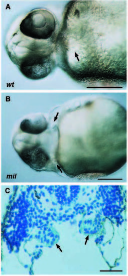
Two separate hearts are formed in the cardia bifida mutations. (A) wild type, ventral view; (B) milm93, ventral view; (C) milm93, histological section; all at 36 hpf. The two hearts (arrows, although one is slightly out of focus) are visible in whole embryos (B) as well as in cross sections (C). Endocardium not shown in these sections. Scale bars, 250 µm for A and B; 100 µm for C. |
Expression Data
Expression Detail
Antibody Labeling
Phenotype Data
| Fish: | |
|---|---|
| Observed In: | |
| Stage: | Prim-25 |
Phenotype Detail
Acknowledgments
This image is the copyrighted work of the attributed author or publisher, and
ZFIN has permission only to display this image to its users.
Additional permissions should be obtained from the applicable author or publisher of the image.
Full text @ Development