
Fig. S4
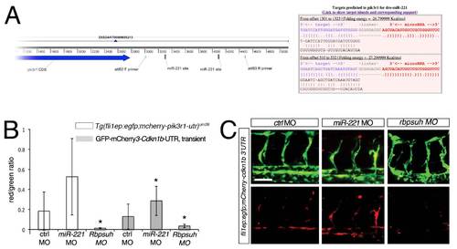
pik3r1 and cdkn1b are targets of miR-221 in zebrafish. (A) Left, pik3r1 transcript showing location of primers used to construct the pik3r1-3′UTR sensor fragment and location of predicted miR-221 binding sites. Right, output from rna22 showing pairing between mature zebrafish miR-221 and putative target sites in the pik3r1 3µ UTR. (B) Quantification of fluorescence in endothelial cell autonomous sensor assay. Red/green ratio indicates comparison of relative voxel intensities derived from 3-dimensional reconstructions of confocal micrographs of either stable Tg(fli1ep:egfp;mcherry-pik3r1-utr)um28 embryos or wild type embryos transiently co-injected with the cdkn1b endothelial sensor construct and Tol2 transposase mRNA. In both cases, embryos were injected with 10 ng control MO, 10 ng mir-221 MO, or 2.5 ng rbpsuh MO. * p<0.05; 7 to 13 embryos for each manipulation, obtained from 3 independent experiments. (C) Confocal micrographs of embryos transiently co-injected with the fli1ep:egfp;mcherry-cdkn1b-utr construct with indicated MOs (doses noted above). Lateral views, dorsal is up, anterior to the left. Scale bar is 50 μm. |
Reprinted from Developmental Cell, 22(2), Nicoli, S., Knyphausen, C.P., Zhu, L.J., Lakshmanan, A., and Lawson, N.D., miR-221 Is Required for Endothelial Tip Cell Behaviors during Vascular Development, 418-429, Copyright (2012) with permission from Elsevier. Full text @ Dev. Cell