
FIGURE
Fig. S2
- ID
- ZDB-FIG-120216-90
- Publication
- Wu et al., 2011 - Ryanodine receptors, a family of intracellular calcium ion channels, are expressed throughout early vertebrate development
- Other Figures
- All Figure Page
- Back to All Figure Page
Fig. S2
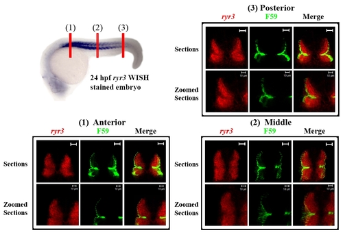
ryr3 mRNA expression is confined to the fast muscle fibres throughout the myotome at 24 hpf. Double fluorescent cross-sections (1-3) were prepared by labelling a 24 hpf zebrafish embryo with a fluorescence substrate (i.e. FastRed; red) for ryr3 in situ hybridisation and the F59 antibody (green) for immunostaining. The position of cross-sections 1-3 are illustrated in the 24 hpf ryr3 WISH labelled embryo (top left) which has been stained with BM purple and laterally orientated with anterior to the left. Scale bars = 20 μm, unless otherwise indicated. |
Expression Data
Expression Detail
Antibody Labeling
Phenotype Data
Phenotype Detail
Acknowledgments
This image is the copyrighted work of the attributed author or publisher, and
ZFIN has permission only to display this image to its users.
Additional permissions should be obtained from the applicable author or publisher of the image.
Full text @ BMC Res. Notes