
Fig. 2
- ID
- ZDB-FIG-120130-2
- Publication
- Schuhmacher et al., 2011 - Evolutionary relationships and diversification of barhl genes within retinal cell lineages
- Other Figures
- All Figure Page
- Back to All Figure Page
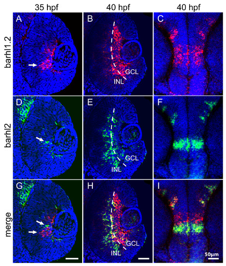
Double fluorescent in situ hybridization of barhl1.2 and barhl2. Confocal sections through the central retina (A-B, D-E and G-H) or diencephalon (C, F and I) of embryos hybridized with both barhl1.2 and barhl2 antisense RNA probes. Stages analyzed are indicated. Nuclei were stained with DAPI (blue). All pictures represent a frontal view (anterior is always to the top). (A-C) barhl1.2 RNA antisense probe revealed with Cy3 (red). (D-F) barhl2 RNA antisense probe revealed with FITC (green). (G-I) green and red channel merged. White arrows in (A, D and G) indicate non-overlapping expression of the two genes. Dashed line in (B, E and H) highlights the boundary between the ganglion cell layer (GCL) and the inner nuclear layer (INL). |
| Genes: | |
|---|---|
| Fish: | |
| Anatomical Terms: | |
| Stage Range: | Prim-15 to Prim-25 |