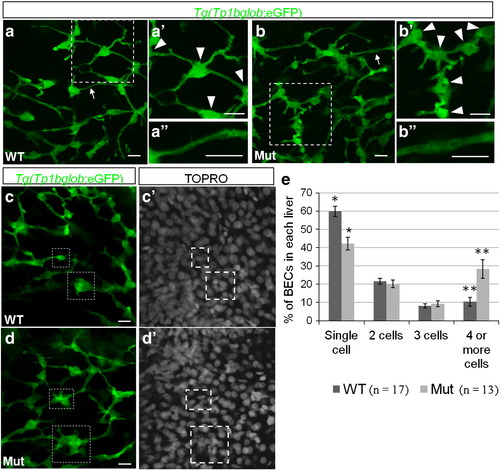
The intrahepatic biliary network is disorganized in cldn15lbfh290 mutant livers. a–d, Whole-mount confocal projections of Tg(Tp1bglob:eGFP)um14 wildtype and mutant livers at 100 hpf. a–b, The biliary network in the mutant larvae appears to be disorganized. a′–b′, Magnification of the boxed regions. In the mutant livers, the cell bodies of the BECs (arrowheads) are closer together, and thus the ducts are shorter. a′′–b′′, Magnification of the duct labeled by the arrow. In the mutant livers, the ducts appear dilated. c–d, At 100 hpf, a majority of the BECs present as individual cells in wildtype livers. In the mutant livers, multiple BECs are clustered together at this stage (boxed regions). e, Percentage of BECs that present as single cells, doubles, triples, or in clusters of four or more cells. Wildtype livers have almost double the number of single cells and almost 3 times less cells in clusters of 4 or more cells (* p = 0.00056, ** p = 0.006). Error bars represent SEM. All scale bars are 10 μm.
|
