
Fig. S4
- ID
- ZDB-FIG-111215-43
- Publication
- Phillips et al., 2011 - Harmonin (Ush1c) is required in zebrafish Muller glial cells for photoreceptor synaptic development and function
- Other Figures
- All Figure Page
- Back to All Figure Page
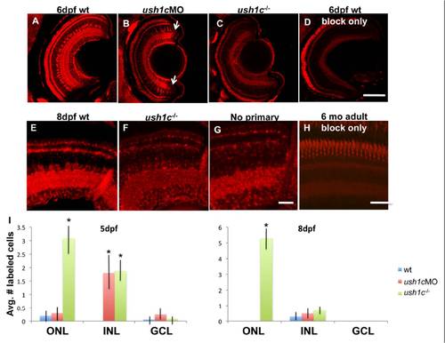
Anti-zebrafish harmonin localization in 6 and 8dpf retinas. (A) Bright, even labeling is seen in Müller glial cells throughout the retina in 6d wild-type animals. (B) Müller cells in 6d morphant animals are faintly labeled in the central retina, but more brightly labeled adjacent to the ciliary marginal zones (arrows). (C) No Müller cell labeling is observed in stagematched ush1c homozygous mutant retinas. (D) A stage matched wild-type embryo treated with blocking solution provides a control for autofluorescence of the photoreceptor outer segments and plexiform layers. (E) Harmonin in 8dpf retinas shows persistent staining in Müller cells and no detectable photoreceptor staining above background levels. Harmonin localization in Muller cells of 8dpf mutants is diminished (F) to background levels. Panel G shows an 8d retina processed with the ABC and TSA kits without incubation in primary antibody to demonstrate the nonspecific signal from synaptic and photoreceptor outer segment membranes. Panel H shows autofluorescence in a 6 mo adult retina incubated with blocking solution only. All negative control images were captured with the same exposure settings as the experimental panels to which they are compared. (I): graphs of retinal cell death by cell layer in 5 and 8dpf larvae. Asterisks denote statistical significance. Scale bars: A-D, H 50 μm; E-G, 10 μm |