|
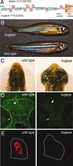
Loss of Lrp2 expression in bugeye mutants. A: Structure of Lrp2 indicating amino acid changes in bugeye and 5cben lines. B, C: Appearance of adult wild type and bugeye zebrafish in lateral (B) and dorsal (C) views highlighting the large eye phenotype in mutants. D: Immunohistological detection of Lrp2 (green) in retinal pigment epithelium (arrow) and ventricular system (arrowhead) in wild type but not in bugeye embryos at 48 hpf using polyclonal antisera directed against multiple epitopes in the extracellular domain of the receptor polypeptide. E: Loss of Lrp2 expression (red) in immunohistological sections of the pronephros in 4-dpf-old bugeye larvae. White lines indicate the position of the pronephric ducts. Complete loss of Lrp2 was also documented in 5cben (data not shown).
|