
Fig. 6
- ID
- ZDB-FIG-111028-3
- Publication
- Chang et al., 2011 - Betanodavirus induces oxidative stress-mediated cell death that prevented by anti-oxidants and zfcatalase in fish cells
- Other Figures
- All Figure Page
- Back to All Figure Page
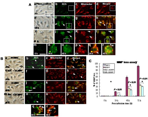
Identification of RGNNV-induced ROS production and the effect of ROS on mitochondrial morphology and loss of ΔΨ in GF-1 cells. Phase-contrast and fluorescence micrographs showing ROS production (the Image-iT LIVE Green Reactive Oxygen Species Detection Kit) and mitochondrial morphology (stained by Mito tracker) were in the same cells. (A) RGNNV-infected GF-1 cells at 0 h (a–d), 24 h (e–h; ROS produced in cells, and 48 h (i–l). The elongated mitochondrial network at 0 h in A:d is indicated by arrow in A:p. ROS production at 0 h in A:m is indicated in open square in A:b; at 24 h pi in A:n and p is indicated open square in A:f and h; at 48 h pi in A:o and r is indicated in open square in A:j and l. Breakdown of mitochondrial fission (indicated by arrows) at 48 h pi in A:r is indicated open square in A:l. Scale bar = 10 μm. (B) RGNNV-infected GF-1 cells treated with NAC at 24 h (e–h) and 48 h pi (m–p), or not treated at 24 h (a–d) and 48 h pi (i–l) with RGNNV infection. Blockade of mitochondrial breakdown in RGNNV-infected GF-1 cells at 48 h pi in B:p is indicated open square in B:l, which were appeared some dot of mitochondria and indicated by arrow; without RGNNV-infected cells at 48 h pi in B:r is indicated open square in B:p, which have shown more longer mitochondria in length that indicated by arrow. Scale bar = 10 µm. (C) The effect of anti-oxidants NAC and DPI on ΔΨ in cells infected with RGNNV. The ΔΨ (MMP loss) of RGNNV-infected GF-1 cells treated or not treated with NAC or DPI was determined at 0, 24, 48, and 72 h pi in triplicate. Statistical comparisons were made using either a paired or unpaired Student′s t-test, as appropriate. *P<0.05. |